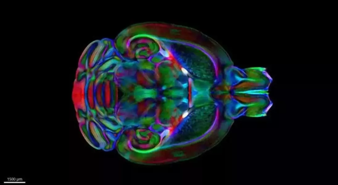
A groundbreaking advancement in neuroscience has emerged with the introduction of the Duke Mouse Brain Atlas, a comprehensive three-dimensional mapping tool developed collaboratively by researchers at Duke University School of Medicine, the University of Tennessee Health Science Center, and the University of Pittsburgh. This atlas integrates advanced imaging techniques such as MRI, microCT, and light sheet microscopy to provide an unprecedented level of detail, from macroscopic brain structures down to individual cells. By eliminating imaging distortions, it establishes a standardized reference framework that enhances precision in measuring changes in brain structure and facilitates data sharing among scientists studying neurodegenerative diseases like Alzheimer’s and Huntington’s.
According to G. Allan Johnson, a distinguished professor of radiology at Duke University, this marks the first truly three-dimensional stereotaxic atlas of the mouse brain. The term "stereotaxic" signifies that the atlas accurately reflects the brain's appearance in living organisms, complete with external landmarks that guide experimental procedures. The development of this atlas addresses challenges posed by various imaging methods, which often produce high-resolution views but introduce distortions during tissue preparation and scanning, complicating comparisons across studies.
The creation process involved multiple stages. Initially, the team utilized MRI with diffusion tensor imaging to capture detailed three-dimensional images of five postmortem mouse brains at an extraordinary resolution of 15 microns—2.4 million times higher than clinical MRIs. Subsequently, these images were merged with microCT scans of the mouse skull to identify critical bony landmarks. Finally, the researchers employed light sheet microscopy on extracted brains to map cellular details within the same spatial framework. This combination of techniques provides one of the most comprehensive maps of the mouse brain ever devised.
Beyond its technical sophistication, the Duke Mouse Brain Atlas is freely accessible through open-source display packages, making it a valuable resource for both experts and laypeople. Grade school students can marvel at the intricate beauty of the brain, while neuroscientists benefit from obtaining more accurate measurements of brain changes. Current applications include tracking neurodegeneration in mouse models affected by Alzheimer’s disease, Huntington’s disease, and exposure to toxic substances such as metals and pesticides.
This innovative tool not only accelerates research into neurological disorders but also democratizes access to cutting-edge brain mapping technology. By providing a common spatial framework, it enables seamless integration of molecular, structural, and functional data across studies, paving the way for transformative discoveries in neuroscience.
